
What Are the Common Causes of Sleep Apnea?
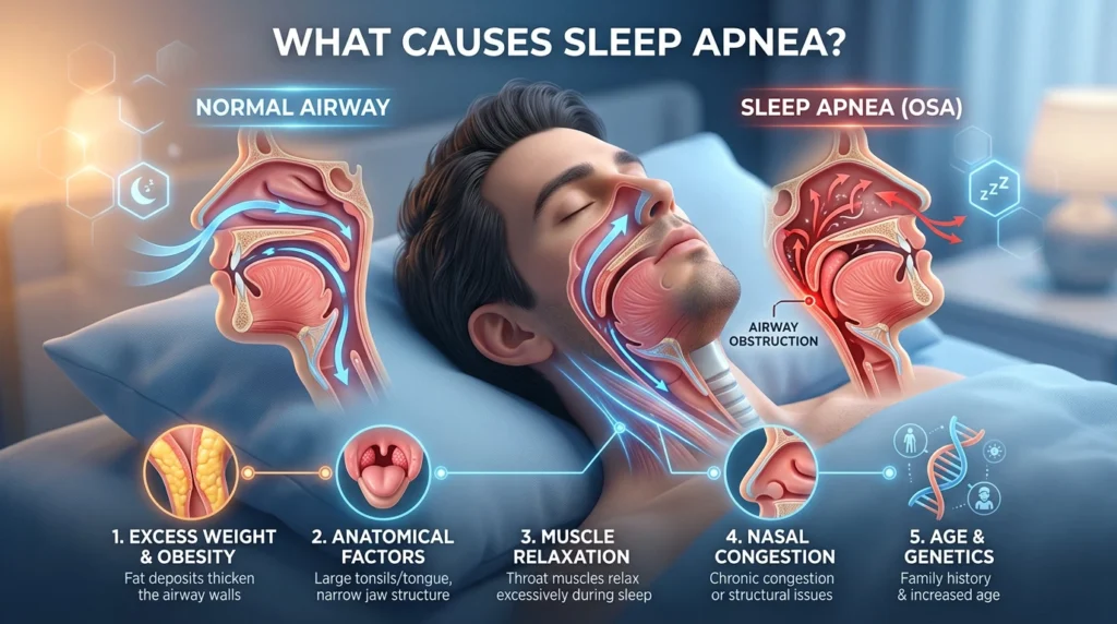
What causes sleep apnea? Sleep apnea is not one disease with one cause: it is a group of disorders in which breathing repeatedly stops or becomes severely restricted during sleep. A person can be young, physically fit and still have sleep apnea. Another can develop it after gaining weight, entering menopause or beginning long-term opioid treatment.
Understanding which type is occurring is important because an obstructed throat, an unstable respiratory-control system and medication-related breathing suppression are not the same problem. They may produce similar symptoms, but they do not always require the same test or treatment.
Obstructive Sleep Apnea Begins with a Collapsing Airway
Obstructive sleep apnea, or OSA, is the most common type. It occurs when the upper airway repeatedly narrows or closes even though the chest and diaphragm continue trying to breathe. Airflow falls, blood-oxygen levels may decline and carbon dioxide rises. The brain then produces a brief arousal that restores muscle activity and reopens the airway.
That arousal may last only a few seconds. The sleeper often has no memory of it, even when the cycle happens dozens of times an hour.
The vulnerable section of the airway lies behind the tongue and soft palate. Unlike the trachea, this portion has no rigid cartilage holding it permanently open. Its size depends partly on surrounding anatomy and partly on the activity of muscles that stiffen and enlarge the airway. Muscle activity normally decreases during sleep. In a person with a narrow or unusually collapsible airway, that normal reduction can be enough to stop airflow.
The Role of the Tongue, Jaw and Throat
There is rarely a single offending structure. MRI research has found that people with OSA may have larger tongues, thicker lateral pharyngeal walls and greater total soft-tissue volume around the airway. Fat may also accumulate within the tongue itself.
The shape of the facial skeleton can contribute independently of body weight. A small or recessed lower jaw leaves less room behind the tongue. A narrow upper jaw, elongated soft palate or unusually positioned hyoid bone can further reduce the available airway. These anatomical features help explain why OSA sometimes runs in families and why a slim adult can have severe apnea.
In children, enlarged tonsils and adenoids are common contributors. A child with obstructive apnea may snore, breathe through the mouth or sleep restlessly, but may display hyperactivity and difficulty concentrating rather than obvious daytime sleepiness. Craniofacial conditions, including Down syndrome, can add several anatomical risk factors at once.
Chronic nasal obstruction from allergies, a deviated septum or swollen nasal tissue does not necessarily cause OSA by itself. It can, however, increase breathing resistance, encourage mouth breathing and make an already vulnerable airway harder to stabilize.
Physiology Alone Doesn’t Always Cause Sleep Apnea
One of the more surprising facts about obstructive sleep apnea is that a person can have a throat that is anatomically prone to collapse yet breathe normally because the tongue muscles are working unusually hard—even while awake. Research measuring activity in the genioglossus, the principal muscle that pulls the tongue forward, found that people with Obstructive Sleep Apnea use more compensatory muscle activity than people without it.
When sleep begins, that compensation diminishes, exposing the unstable airway underneath. In other words, the airway does not necessarily become defective at bedtime; sleep removes part of the muscular support that had been concealing the defect all day. This helps explain why examining an awake patient’s throat cannot reliably establish whether they have OSA or how severe it is: the failure becomes visible only when anatomy, muscle control and sleep interact. (Mezzanotte et al., Journal of Clinical Investigation)
Why Body Weight Raises the Risk
Excess body weight is an important risk factor, but the explanation is more complex than fat simply “blocking the throat.” Tissue deposited around the tongue, neck and pharynx can make the airway smaller and more compressible. Abdominal fat can also reduce lung volume, particularly when a person lies down. Lower lung volume provides less downward traction on the upper airway, allowing it to collapse more readily.
Weight changes can affect severity. A person whose snoring becomes much louder after gaining 25 pounds may have crossed from a vulnerable but functional airway into clinically significant obstruction. Weight loss often reduces the number of respiratory events, but it does not reliably cure OSA because jaw structure, soft-tissue anatomy and respiratory-control characteristics remain.
The National Heart, Lung, and Blood Institute identifies obesity, large tonsils and hormonal changes among the factors that can narrow the upper airway. None is present in every patient.
Anatomy Does Not Explain Every Case
Two people with similarly narrow throats can produce very different sleep-study results. Researchers have identified several physiological traits that influence whether a narrowing airway becomes repeated apnea.
Some people’s upper-airway muscles do not respond strongly or quickly enough when breathing becomes restricted. Others have unstable ventilatory control, meaning that the body overcorrects after a disturbance and then allows breathing effort to fall too far. Researchers call this tendency “high loop gain.”
The brain’s arousal threshold also plays a role. If a person wakes too easily in response to minor airway narrowing, sleep repeatedly fragments before the respiratory system can stabilize. If the threshold is unusually high, an obstructive event may last longer before the brain intervenes. A major review of sleep-apnea pathophysiology describes how airway anatomy, muscle responsiveness, arousal and breathing control interact.
This combination of traits is one reason OSA severity cannot be predicted accurately from neck size, weight or snoring volume alone.
Alcohol, Sedatives and Sleeping Position
Alcohol usually does not create OSA in a person with a completely stable airway, but it can expose or aggravate an existing vulnerability. It reduces upper-airway muscle activity and may delay the arousal that ends an obstruction. Sedatives and some sleep medications can also worsen breathing in susceptible patients, although their effects differ by drug and individual physiology.
Sleeping on the back allows gravity to move the tongue and soft palate toward the rear of the throat. Some people therefore have positional OSA: their apnea-hypopnea index is considerably higher while supine than while sleeping on either side. A study that records body position can identify this pattern.
Smoking is associated with OSA as well, potentially through inflammation and fluid accumulation in the upper airway. It should be treated as a risk factor rather than proof of causation in an individual patient.
Age, Sex Hormones and Pregnancy
OSA becomes more common with age. Changes in tissue elasticity, fat distribution, muscle responsiveness and sleep architecture may all contribute.
Men develop OSA more often during early and middle adulthood, but women’s risk rises after menopause. The increase is not explained entirely by weight. Changes in estrogen and progesterone may affect fat distribution, ventilatory control and upper-airway muscle function. Women are also more likely to report insomnia, fatigue or morning headaches instead of the stereotypical complaint of falling asleep during the day, which can delay recognition.
Pregnancy can temporarily increase risk through weight gain, fluid shifts, nasal congestion and changes in upper-airway dimensions. New loud snoring, witnessed breathing pauses or significant daytime impairment during pregnancy should be discussed with a clinician rather than dismissed as ordinary pregnancy fatigue.
What Causes Central Sleep Apnea?
Central sleep apnea, or CSA, works differently. The airway may remain open, but respiratory effort stops because the brain’s breathing-control system does not maintain a stable signal to the respiratory muscles.
Heart failure can produce a characteristic waxing-and-waning pattern called Cheyne-Stokes respiration. Delayed circulation and heightened sensitivity to carbon dioxide contribute to cycles of overbreathing, falling carbon-dioxide levels and temporary pauses in respiratory effort.
Opioids can suppress brainstem respiratory drive and cause central apneas, slow breathing or irregular breathing. The risk is particularly relevant with long-acting drugs and higher doses, although no dose can be assumed safe for every patient.
Central apnea can also occur after a stroke, with certain neurological or neuromuscular diseases, or during high-altitude exposure. At altitude, low oxygen stimulates rapid breathing. Carbon dioxide may then fall below the level required to sustain breathing during sleep, producing alternating periods of apnea and recovery. The Mayo Clinic’s overview of central sleep apnea describes these cardiac, neurological, medication-related and altitude-associated mechanisms.
Some patients develop central events after CPAP successfully removes their obstructive events. This is called treatment-emergent central sleep apnea. It often resolves with continued treatment, but it requires clinical follow-up rather than an improvised change in pressure settings.
A Sleep Test Shows Which Mechanism Is Present
Snoring cannot reveal whether respiratory effort continued during a breathing pause. Neither can a smartwatch or a stand-alone pulse oximeter. A medically interpreted sleep study examines patterns of airflow, breathing effort, oxygen saturation and heart rate; laboratory polysomnography records additional signals such as brain activity and sleep stage.
An at-home sleep apnea test may be appropriate for an adult whose history suggests uncomplicated OSA. Suspected central apnea, opioid-related breathing abnormalities, significant heart or lung disease, neuromuscular weakness or unexplained sleep symptoms may require an attended laboratory study.
The objective is not simply to confirm that breathing stops. It is to establish why it stops, how often it happens and what physiological effects follow—information that determines which treatment has a realistic chance of working.
Frequently Asked Questions
Obstructive sleep apnea occurs when the muscles supporting the tongue and throat relax during sleep, allowing a vulnerable upper airway to narrow or close repeatedly.
No. Excess weight increases the risk, but people at any weight can develop sleep apnea because of jaw structure, tongue size, enlarged tonsils, hormonal changes or poor upper-airway muscle responsiveness.
Alcohol can worsen or reveal obstructive sleep apnea by relaxing the throat muscles and delaying the body’s response to an obstruction. Its effect is usually greatest when consumed near bedtime.
Risk can run in families. Inherited features such as facial structure, jaw size, fat distribution and the anatomy of the upper airway may make apnea more likely, although having an affected relative does not guarantee that you will develop it.
Central sleep apnea occurs when the brain temporarily fails to maintain the signal that drives breathing. Possible causes include heart failure, opioid use, stroke, certain neurological conditions and high-altitude exposure.
It may become noticeable after weight gain, menopause, tonsil enlargement, increased alcohol or sedative use, opioid treatment or the development of heart or neurological disease. In many cases, an existing airway vulnerability has gradually worsened rather than appearing overnight.
Why can sleep apnea appear suddenly?
It may become noticeable after weight gain, menopause, tonsil enlargement, increased alcohol or sedative use, opioid treatment or the development of heart or neurological disease. In many cases, an existing airway vulnerability has gradually worsened rather than appearing overnight.